Powered By Blogger

Sunday 15 November 2015

DISEASES OF REPRODUCTIVE SYSTEM

DISEASES OF FEMALE REPRODUCTIVE SYSTEM


  1. PELVIC INFLAMMATORY DISEASE-  
  • Inflammation of the female reproductive organs often dut to a sexually transmitted infection.
  • Most common between the ages of 15 to 24.
  • Pelvic inflammatory disease is a common cause of pain in the pelvic region in women.
  • The infection spreads upwards from the vagina to the uterus and fallopian tubes.
  • Also takes place due to insetion of intra-uterine contraceptive device.
SYMPTOMS:
  •  Pain in pelvic region.
  • Fever
  • An abnormal vaginal discharge.
  • Heavy or prolonged periods.
  • Pain during sexual intercourse
  • Tiredness
  • nausea, vomiting 
  • damaged fallopian tubes 


pelvic inflammatory disease 


video to understand pelvic inflammatory disease
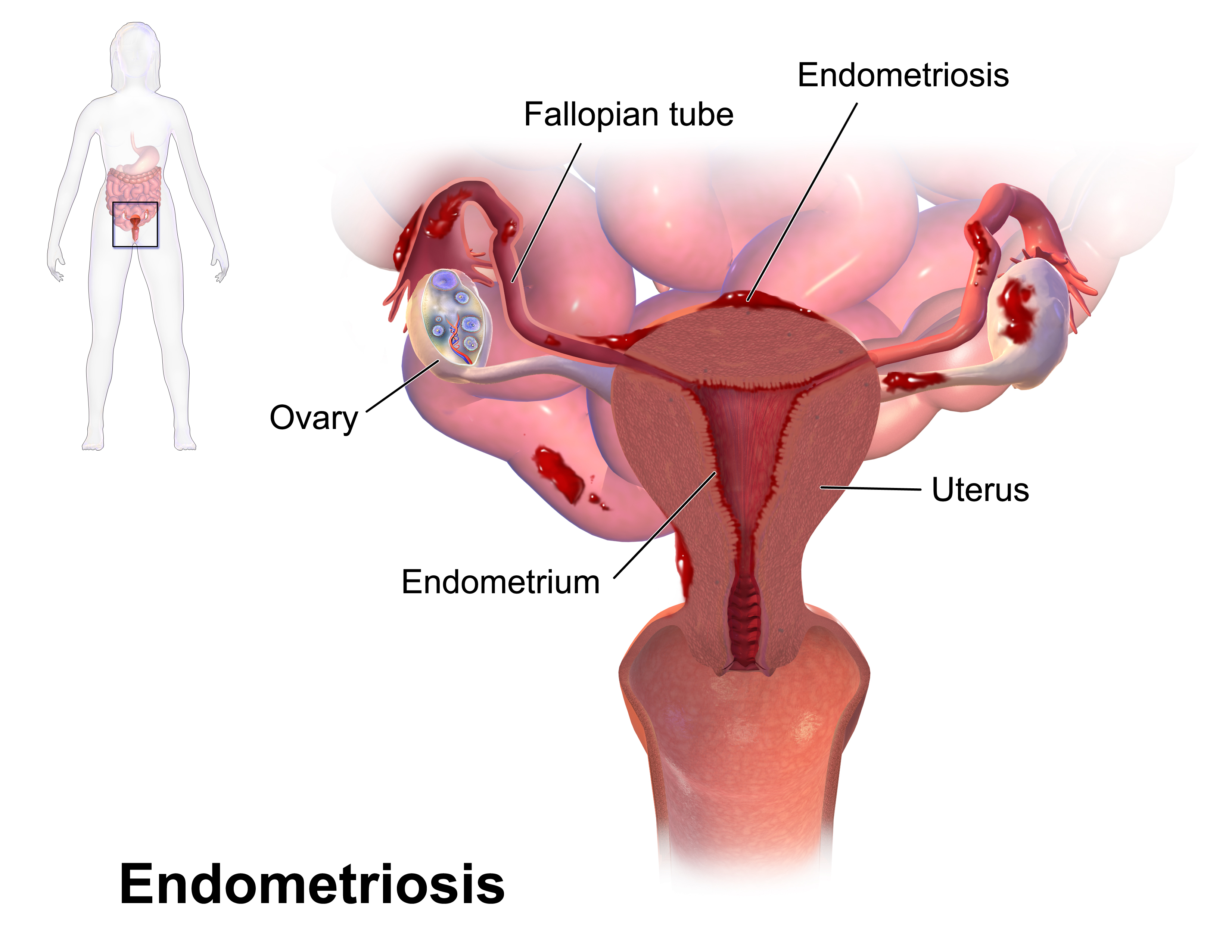

2. ENDOMETRIOSIS-  
  • A condition in which endometrial tissue, which lines the uterus but in this case it is attached to other organs in the abdomen.
  • Common between the ages of 30 and 45.
  • The lining of the uterus, known as endometrium , is normally shed once a month during menstruation and then regrows.
  • in endometrium, some pieces of the lining are attached to organs in the pelvic cavity, such as the ovaries and the lower intestine.
  • These pieces of endometrial tissue react to the hormones of the menstrual cycle and bleed during menstruation.
  • The blood cannot leave the body through the vagina and this can cause irritation of the surrounding tissues, leading to pain in the abdomen and eventually scarring.
  • irritation of the ovaries may lead to blood filled cysts.
  • it is a common condition, affecting as many as 1 in 5 women of childbearing age.
  • Women who do not have children until they are in their 30s and those who remain childless are more likely to develop the condition.
SYMPTOMS- 

  • Pain in lower abdomen, which often becomes more severe just before and during menstrual periods.
  • Heavier menstrual bleeding
  • Pain during sexual intercourse
  • If endometrium grows on the lower intestine, you may develop diarrhoea or constipation, pain during bowel movements, and in rare cases, bleeding from the rectum during menstruation.


video for endometriosis 


POLYCYSTIC OVARY SYNDROME - 

  • Multiple , small fluid  filled cysts in the ovaries associated with a sex hormone imbalance.
  • Affects females of childbearing age.
  • Sometimes runs in families.
  • Many women have multiple fluid filled cysts in their ovaries but most of these women do not have polycystic ovary syndrome, which is the presence of multiple ovarian cysts associated with an imbalance of the sex hormones and certain other characteristics , such as acne , excessive body hair, and menstrual irregularities. 
  • Higher levels of male sex hormone testosterone, may prevent ovulation, thus reducing fertility.
  • The underlying cause of PCOS cause is increased resistance of body tissues to be hormone insulin that is a feature of the syndrome and is thought to play an important part.
  • To compensate for the increased insulin resistance, the pancreas produces excessive insulin, ehich in turn, may lead to over production of testosterone , high levels of which disrupt normal functioning of the ovaries.
SYMPTOMS- 
  • Infrequent or absent periods 
  • Obesity 
  • excessive hair growth on the face, around the nipples or on the lower abdomen.
  • Thinnig of the hair on the head.
  • Long lasting acne.
  • Women with PCOS have an increased risk of diabetes mellitus, high blood pressure, coronary heart disease and cancer of the uterus.


video for PCOS 


FIBROIDS-   

  • Common, noncancerous tumours that grow slowly within the muscular wall of the uterus.
  • Most common between the ages of 35 to 55.
  • fibroids are non-malignant growths in the uterus that consist of muscular and fibrous tissue.
  • Small Fibroids may not cause problems but larger ones may affect menstrution or fertility.
  • Fibroids occur singly or in groups and may be small as a pea or as big as a grapefruit.
SYMPTOMS- 
  • Prolonged menstrual bleeding 
  • Abdominal pain during periods
  • Heavy bleeding during periods.

  • Heavy blood loss may lead to anaemia causing pale skin and tiredness.
  • Large fibroids may distort the uterus which can sometimes result in infertility and possibly in recurrent miscarriages.
  • During pregnancy a large fibroid may cause the foetus to lie in an abnormal position.
  • Fibroids may also press on the bladder causing a need to pass urine often, or on the rectum, causing back pain.


Video for fibroids 


CERVICAL INTRAEPITHELIAL NEOPLASIA -  

  • Changes in the surface cells of the cervix that may become cancerous.
  • Most common between the ages of 25 to 35.
  • There are 3 grades, depending on the severity of the changes in the abnormal cells; mild, moderate and severe.
  • Mild CIN returns to normal state by itself but moderate or severe CIn may progress to the concer of cervix, if not treated.
  • It is caused usually due to exposure to some types of human papillomavirus.
  • Other risk factors for the development of CIN include unprotected sex at an early age and unprotected sex with many partners because these activities are associated with an increased risk of acquiring HPV infection.


VULVOVAGINITIS- 

  • Inflammation of the vulva and vagina, causing itching and soreness.
  • There may also be pain during sexual intercourse and a discharge from the vagina.
  • Most cases of vulvovaginitis are caused by an infection, either with the fungus Candida albicans, or the protozoan Trichomonas vaginalis, which is the cause of the sexually transmitted infection trichomoniasis.
  • Women with diabetes mellitus have an increased risk of fungal vulvovaginits.
  • The condition may be caused by irritation from perfumed bath products, detergents, deoderants, or contraceptive creams.


video of vulvovaginitis 


DISEASES OF MALE REPRODUCTIVE SYSTEM
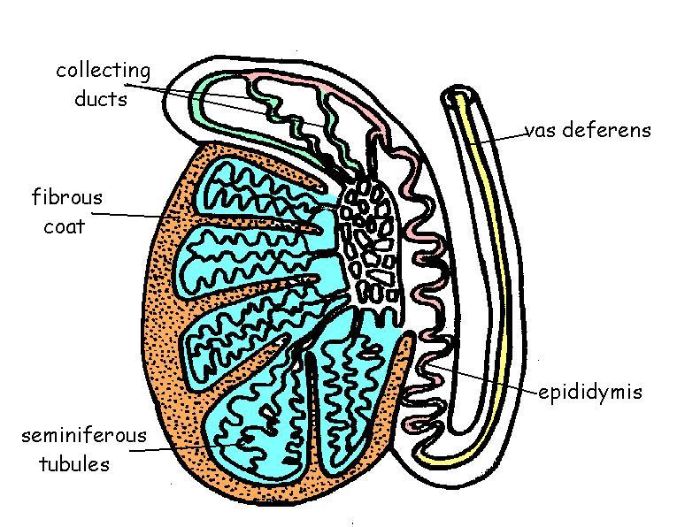
Epidermal cysts -  

  • More common over the age of 40.
  • Epididymal cysts or spermatoceles, are harmless fluid - filled sacs that form in the epididymis, which stores and transports sperm away from the testis.
  • Small epididymal cysts are common, particularly in men over the age of 40.
  • The cysts develop slowly and are usually painless.
  • In many cases, there are multiple cysts, which can be felt as distinct, painless swellings like a tiny bunch of grapes on top of and behind the testis.


Torsion of the testis - 

  • Twisting of the testis within the scrotum, causing severe pain.
  • Most common between the ages of 12 and 18 but can occur at any age.
  • Each testis is suspended in the scrotum on a spermatic cord.
  • The spermatic duct and the blood vessels that supply the testis are contained in the spermatic cord.
  • If the spermatic cord becomes twisted, the flow of blood to the testis is restricted, causing severe pain in the scrotum.
  • Torsion of the testis usually affects only one of the testes.
  • if sometimes occurs often strenous activity but may develop for no apparent reason, even during sleep.
  • Torsion of the testis most commonly occurs during adolescence, but it can develop at any age.
  • The condition is potentially serious , torsion may result in permanent damage to the testis if not treated immediately.
SYMPTOMS-

  • Sudden pain in the scrotum that tends to increase in severity.
  • Pain in the groin and lower abdomen.
  • Redness and extreme tenderness of the scrotum on the affected side.
  • The severity of pain can cause nausea and vomiting.


BALANITIS - 

  • Inflammation of the head of the penis and foreskin.
  • More commin in children.
  • In addition, there may be a discharge , and a rash may develop.
  • The disorder can be caused by a bacterial infection, a fungal infection such as thrush or an allergic reaction.
  • It may also be due to a sexually transmitted infection such as the protozoal infection trichomoniasis.
  • A tighter than normal foreskin may increase the risk of infection by preventing effective cleaning of the glans.
  • Men with diabetes mellitus are more susceptible to the condition because their urine contains high level of glucose, which can encourage growth of micro organisms.
  • This leads to infection and inflammation at the opening of the urethra.
  • Excessive use of antibiotics can increase the risk of a fungal infection by temporarily lowering the body's natural defences against this type of infection.
  • The condition may also occur as a result of sensitivity of the penis to certain chemicals, such as those found in some condoms, contraceptive creams, detergents and washing powders.

BENIGN PROSTATIC HYPERTROPHY - 

  • It is characterized by proliferation of the cellular elements of the prostate.
  • Chronic bladder outlet obstruction secondary to BPH may lead to urinary retention, renal insufficiency, recurrent urinary tract infections, gross hameturia, and bladder calculi.
SYMPTOMS - 
  • Urinary frequency 
  • urinary urgency 
  • Hesitancy
  • Incomplete bladder emptying
  • Straining
  • Decreased force of stream
  • Dribbling


* Do Give your suggestions *

Thamkyou 


Friday 13 November 2015

MALE REPRODUCTIVE SYSTEM

SCROTUM -  The scrotum is a sac like organ made of skin and muscles that houses the testes.
It is located inferior to the penis in the pubic region.
The scrotum is made up of 2 side by side pouches with a testis located in each pouch.
The smooth muscles that make up the scrotum allow it to regulate the distance between the testes and the rest of the body.
When the testes become too warm to support spermatogenesis, the scrotum relaxes to move the testes away from the body's heat.
Conversely, the scrotum contracts to move the testes closer to the body's core heat when temperature drop below the ideal range for spermatogenesis.

TESTES-  The 2 testes, also known as testicles, are the male gonads responsible for the production of sperm and testosterone.
The testes are ellipsoid glandular organs around 1.5 to 2 inches long and an inch in diameter.
Each testis is found inside its own pouch on one side of the scrotum and is connected to the abdomen by the spermatic cord and cremaster muscle.
The cremaster muscles contract and relax along the scrotum to regulate the temperature of the testes.
The inside of the testes is divided into small compartments known as lobules.
Each lobule contains a section of seminiferous tubule lined with epithelial cells.
These epithelial cells contain many stem cells that divide and form sperm cells through the process of spermatogenesis.

EPIDIDYMIS-  The epididymis is a sperm storage area that wraps around the superior and posterior edge of the testes.
The epididymis is made up of several feet long, thin tubules that are tightly coiled into a small mass.
Sperm produced in the testes move into the epididymis to mature before being passed on through the male reprouctive organs.

The length of the epididymis delays the release of the sperm and allows them time to mature.


SPERMATIC CORDS AND DUCTUS DEFERENS -  Within the scrotum , a pair of spermatic cords connects the testes to the abdominal cavity.
The spermatic cords contain the ductus deferens along with nerves, veins, arteries and lymphatic vessels that support the function of the testes.
The ductus deferens, also known as vas deferens, is a muscular tube that carries sperm superiorly from the epididymis into the abdominal cavity to the ejaculatory duct.
The ductus deferens is wider in diameter than the epididymis and uses its internal space to store mature sperm.
The smooth muscles of the walls of the walls of the ductus deferens are used to move sperm towards the ejaculatory duct through peristalsis.

Structure of testis 


SEMINAL VESICLES -  Pair of glands that store and produce some of the liquid portion of semen.
The seminal vesicles are about 2 inches in length and located posterior to the urinary bladder and anterior to the rectum.
The liquid produced by the seminal vesicles contains proteins and mucus and has an alkaline pH to help sperm survive in the scidic environment of the vagina.
The liquid also contains fructose to feed sperm cells so that they survive long enough to fertilize the ovum.

EJACULATORY DUCT - The ductus deferens passes through the prostate and joins with the urethra at the structure known as ejaculatory duct.
The ejaculatory duct contains the ducts from the seminal vesicles as well.
During ejaculation, the ejaculatory duct opens and expels sperm and the secretions from the seminal vesicles into the urethra.

URETHRA- Semen passes from the ejaculatory duct to the exterior of the body via the urethra an 8 to 10 inch long muscular tube.
The urethra passes through the prostate and ends at the external urethral orifice located at the tip of the penis.
Urine exiting the body from the urinary bladder also passes through the urethra.

PROSTATE GLAND -  The prostate is a walnut shaped gland that borders the inferior end of the urinary bladder and surrounds the urethra.
The prostate produces a large portion of the fluid that makes up semen.
This fluid is milky white in color and contains enzymes, proteins and other chemicals to support and protect sperm during ejaculation.
The prostate also contains smooth muscle tissue that can construct to prevent the flow of urine or semen.

COWPER'S GLAND -  The cowper's glands is also known as bulbourethral glands, are a pair of pea- sized glands located inferior to the prostate and anterior to the anus.
The cowper's glands secrete a thin alkaline fluid into the urethra that lubricates and neutralizes acid from the urine remaining in the uretra after urination.
This fluid enters the urethra during sexual arousal prior to ejaculation to prepare the urethra for the flow of semen.

PENIS-  The penis is the male external sexual organ located superior to the scrotum and inferior to the umbilicus.
The penis is roughly cylindrical in shape and contains the urethra and the external opening of the urethra.
Large pockets of erectile tissue in the penis allow it to fill with blood and become erect.
The erection of the penis causes it to increase in size and become turgid.
The function of the penis is to deliver semen into the vagina during sexual intercourse.
In addition to its reproductive function, the penis also allows for the excretion of urine through the urethra to the exterior of the body.


MALE REPRODUCTIVE SYSTEM 


Through this video you will understand male reproductive system better 

*thankyou*

*do give your suggestions *


Thursday 12 November 2015

A LIFE INTO A LIFE 


Fertilization and development of foetus 

FERTILIZATION 

 It is the union of human egg and sperm occurring in the fallopian tubes.

 The result of this union is the production of a zygote cell or fertilized egg.

The sperm on entering the female reproductive system releases enzymes from its acrosome region present in the head region that disintegrates the corona radiata and zona pellucida layer of the ovum, thereby out of million sperms, only one sperm fuses with the female pronucleus.

The sperm head containing the nucleus enters the ovum, thereby degenerating the middle piece and the tail region.


* video of fertilization *


IMPLANTATION 


After fertilization, the fertilized egg propels through the tube towards the uterus.

The cells of the zygote divide repeatedly as the zygote moves down the fallopian tube.

The zygote enters the uterus in 3 to 5 days .

In the uterus, the cells continue to divide, becoming a hollow ball of cells called a blastocyst.

Between 5th and 8th day after fertilization, the blastocyst attaches to the lining of the uterus, usually near the top.

This process is called Implantation, is completed by 9th or 10th day.


DEVELOPMENT OF BLASTOCYST

  •  The wall of  blastocyst is one cell thick except in one area, where it is three or four cells thick.
  • The inner cells develop into an embryo and the outer cells burrow into the wall of the uterus and develop into placenta.
  • The placenta produces several hormones that help maintain the pregnancy.
  • The placenta also carries oxygen and nutrients from mother to foetus and waste materials from foetus to mother.
  • Some of the cells from placenta form an outer layer chorion around the developing blastocyst.
  • Other cells develop into an inner layer amnion, which forms the amniotic sac.
  • The amniotic sac fills with an amniotic fluid and expands to envelope the developing embryo, which floats within it.
DEVELOPMENT OF EMBRYO

  •  The next stage in development is the embryo, which develops within the amniotic sac, under the lining of the uterus on one side.
  • This stage is characterized by the formation of most internal organs and external body structures.
  • Most organs form to begin to form about 3 weeks after fertilization, which equals 5 weeks of pregnancy.
  • At this time, the embryo elongates, first suggesting a human shape.
  • Shortly, thereafter, the area that will become the brain and spinal cord begin to develop.
  • The heart and blood vessels begin to develop earlier by about Day 16.


Embryo at 8 weeks 


DEVELOPMENT OF FOETUS AND PLACENTA 

  • At the end of the 8th week after fertilization, the embryo is considered a foetus.
  • During this stage, the structures that have already formed grow and develop.
  • By 12 weeks of pregnancy- the foetus fills the entire uterus.
  • By about 14 weeks - the sex is identified.
  • By about 16-20 weeks - typically the pregnant woman can feel the foetus moving.
  • Women who have been pregnant before typically feel movements about 2 weeks earlier than women who are pregnant for the first time.
  • By about 24 weeks- the fortus has a chance of survival outside the uterus
  • The lungs continue to mature until near the time of delivery
  • The brain accumulates new cells throughout pregnancy and the first year of life after birth.
  • As, the placenta develops, it extends tiny hair like projections into the wall of the uterus.
  • The projections branch and rebranch in a complicated treelike arrangement.
  • This arrangement greatly increases the area of contact between the wall of the uterus and the placenta, so that more nutrients and waste materials can be exchanged.
  • The placenta is fully formed by 18 to 20 weeks but continues to grow throughout pregnancy.
  • At delivery, it weighs about 1 pound.


Pregnancy


PARTURITION
Parturition, also called birth or childbirth, process of bringing forth a child from the uterus or womb.
It takes place after the gestation period of 9 months.


Stages of pregnancy in the mother's womb ..... * this video will help you to understand the stages of foetal development *

*do out your suggestions* 

THANKYOU 


Wednesday 11 November 2015

A LIFE GIVING RISE TO LIFE 

Today's topic deals with the female reproductive system and the developing sexual characters  


FEMALE REPRODUCTIVE SYSTEM 

The female reproductive system consists of a pair of ovaries situated on the either sides of a pear shaped uterus . In continuation of the ovaries are present a tube like structure known as the oviduct or the fallopian tubes. 

The uterus consists of an upper fundus, the body and the lower cervix which continues into vagina , which forms the birth canal of the foetus. 

The lining of uterus consists of outer perimetrium, middle myometrium and inner endometrium . the endometrial lining consists of blood and mucus ( fluid that helps in lubricating the uterus ). 

The external genitilia of the female reproductive system is known as the vulva , which has two openings, one being the urinary orifice and the other being the vaginal orifice. 

Before moving on to the process of fertilization , lets get well versed with the phenomenon which takes place in the female reproductive system in the absence of fertilization .


Female reproductive system


ACCESSORY GLANDS 

  1. Bartholin's gland - These are two pea sized compound recemose glands located slightly posterior or lower to the left and right of the opening of the vagina.
  2. They secrete mucus to lubricate the vagina.
  3. They secrete relatively minute amounts of fluid when a woman is sexually aroused.

 Location of bartholin's gland - It is a part of the external genitilia - vulva 


  1. Mammary glands - A mammary gland is an organ in female mammals that produces milk to feed young offspring.
  2. Mammals get their name from the word mammary.
  3. In humans, the mammary glands are situated in breasts.
  4. A msture mammary gland consists of lobules.
  5. Each lobule has lactiferous ducts that drains into opening in the nipple.
  6. Humans normally have two mammary glands, in each breast.
  7. They are lined by milk secreting cells, which are cuboidal.
  8. Milk is secreted, when the baby gives a suckling stimulus.
 Mammary glands 


EXTERNAL GENITAL ORGAN- VULVA 

  • The vulva consists of the external genital organs of the female mammal.
  • It consists of labia majora, labia minora, clitoris, vestibule, external urethral orifice , opening of vagina and the hymen.
  • The Presence of hymen determines that the female is a virgin.
  • The hymen breaks when there an intercourse in between the opposite sex.
  • The absence of hymen does not necessarily determine that the female has undergone an intercourse, it can also break at the time of major injuries or while playing sports. 
  • Therefore, do not define a female as a virgin or a non- virgin on the basis of the presence or absence of the hymen respectively.                                                                            Anatomy of vulva


* To understand the female reproductive system in a better way you can watch this video * 


MENSTRUAL CYCLE  

Menstrual cycle is a cycle of 28 days which is characterized by vaginal bleeding for 3-5 days of a month.

As females have two ovary , out of which either of the one releases one ovum into the fallopian tubes.

The ovum is ready to fertilize with the sperm and thefore waits for 14 days.

While is ovary comes into the uterus , the endometrial lining thickens , thereby making the uterus ready for the process of attachment or implantation of the developing foetus after the process of fertilization.

But if fertilization of sperm and ovum does not take place within those 14 days , the ovum is shed off from the body along with the endometrial lining which is formed of blood and mucus , hence caharacterized by vaginal bleeding.

In layman's language it is known as periods because its a regular cycle that takes place every month.

Menstrual cycle or periods is a social taboo as yet in India , but it not something extraordinary , but a usual process in females. 

It is a normal process of expelling the unfertilized ovum from the female's body and nothing else.


* A wonderful animation on menstruation .... do watch it ...... to understand the eternal plan of nature *


THANKYOU 

* do give your suggestions *


Tuesday 10 November 2015

Sex Determination In humans

CONTINUATION OF LIFE .....

In today's topic I would cover topics about the mechanism of determination of sex in humans  

Before moving on to the mechanism of sex determination , i would like to make you all familiar to some important terminologies :

  1. Gene- basic unit of heredity 
  2. Somatic cells - All body cells except reproductive cells 
  3. Gamete- reproductive cells 
  4. chromosome - elongate cellular structure composed of DNA and protein, they are vehicles which carry DNA in cells. 
Lets go into the details of chromosomes 


CHROMOSOMES

Chromosomes are special cellular structures which are formed of highly colied structure DNA which consists of genes. Chromosomes contain the genetic material of the cell  which passes on from one generation to next generation. 

Chromosomes are of basic two types :

  • 22 pairs of Autosomes or somatic chromosomes 
  • and 1 pair of Allosomes or Sex chromosomes 
Allosomes or Sex chromosomes are the ones which are involved in the process of determination of sex of an individual and Autosomes are the ones which have their role in the general body functions . 

Sex chromosomes in case of mammals are of two different types :

XX in case of females and XY in case of males 

These chromosomes are present in the reproductive cells , hence they undergo meiotic division .

Females are Homogametic as they have XX chromosomes and males are heterogametic as they have XY chromosomes , that is the human sperm present in males have both X and Y chromosome , Therefore males are responsible for the process of sex determination of the offspring and not the female .

Furthermore, lets go into the details of the structure of sperm in males and ovum in the females.


HUMAN SPERM 


Structure of sperm 


  • The mammalian sperm cell consists of a head, midpiece and a tail . 
  • The head contains the nucleus with densely coiled chromosomes surrounded by an acrosome.
  • The midpiece has a central core of spiral shaped mitochondria.
  • The tail haleps in propellation pf the sperm 

                                                                   HUMAN OVUM 

  • The mammalian ovum consists of  an outer corona radiata and inner zona pellucida.
  • the cytoplasm of the ovum cell is known as ooplasm which shows the presence of a pronucleus.  

Structure of ovum 


INHERITANCE OF XY CHROMOSOMES 

  • During gamete formation, the normal diploid chromosome is halved. This is called the haploid condition.
  • All the eggs of a female have 22+X.
  • A male produces two types of sperms, one type bears 22+X composition and the other 22+Y .
  • Therefore, in many 100 sperms , 50 have Y chromosomes and 50 X chromosomes.
  • Any one of the two types of sperms can fertilize the egg.
  • If a Y bearing sperm fertilizes the egg, the zygote has the 44+XY composition, and the resulting foetus grows to be a boy.
  • When an X- bearing sperm fertilizes the egg, the resulting zygote has the 44+XX composition, resulting into a girl.
  • All the children inherit one X chromosome from the mother.
  • Therefore, sex is always determined by the other sex chromosome that they inherit from the father. 
  • One who inherits the X chromosome of the father is a girl, while one who inherits the Y chromosome of the father is a boy.


This video will help you all to understand the mechanism of sex determination better 
Thankyou 

*kindly give your suggestions *


Monday 9 November 2015

Life - as it continues

LIFE- as it continues to grow 

Cell growth and cell division 


Cell division involves the distribution of identical genetic material , that is the DNA (Deoxyribonucleic acid) to two daughter cells .

Cell division is of two types :
  • Mitosis 
  • Meiosis 
Mitosis is the type of cell division which takes place in general body cells or somatic cells .
Eg- Liver, Bone marrow ,etc.

Meiosis is the type of cell division which takes place in germ cells or reproductive cells 
Eg- Sperm , Ovum 


Before moving ahead to the mechanism of cell division , I would like you all to get Familiar to some basic terminologies:

  • gene - basic unit of heredity, codes for a specific trait 
  • somatic cells - all body cells except reproductive cells
  • gamete - reproductive cells , eg - sperm and ovum 
  • chromosome - elongate cellular structure composed of DNA and                               protein, they are vehicles which carry DNA in cells.
  • diploid - cellular condition where each chromosome type is                              represented in a pair.
  • haploid- cellular condition where chromosome is represented only                  by single chromosome.
  • homologous chromosome - chromosome of the same size and shape which carry the same type of genes.
  • chromatid - one of the two duplicated chromosomes connected at                        the centromere.
  • centromere - region of chromosome where microtubules attach during mitosis and meiosis. 

chromosome structure chromosome diagram 

2n=4Example - an organism is 2n = 4. 

  • Chromosomes 1 & 2 are homologous chromosomes
  • Chromosomes 3 & 4 are homologous chromosomes
  • Chromosomes 1 & 3 came from the mother
  • Chromosomes 2 & 4 came from the father


Now lets move on to the mechanism of cell division, 

Cell division undergoes a series of cyclic phases namely , G1 - S- G2 - M phase thereby calling it  the cell cycle . 

cell cycle


G1 - first gap

S - DNA synthesis (replication)
G2 - second gap
M - mitosis

Diagrammatic representation of the phases of cell cycle .


The stages of cell cycle are divided into 5 stages :


  1. Interphase - It is the resting or non - mitotic portion of the cell                               cycle . It is comprised of G1, S, G2 stages of the cell                           cycle. 
  2. Prophase-  The first stage of mitosis , the chromosomes condense                       and become visible. The centrioles form and move                              towards opposite ends of the cell. The nuclear                                    membrane dissolves . The mitotic spindle forms from                       the centrioles in case of animal cell and microtubules in                     case of plant cell. Spindle fibers from each centriole                           attach to each sister chromatid at the kinetochore.

ProphaseProphase

                                          Prophase


      3. Metaphase - The centrioles complete their migration to the poles. The chromosomes line                                   up in the middle of the cell. 


MetaphaseMetaphase

Metaphase 

4. Anaphase - Spindles attached to kinetochores begin to shorten . This exerts a force on sister                           chromatids that pulls them apart. Spindle fibers continue to shorten, pulling                                 chromatids to opposite poles. This ensures that each daughter cell gets identical                           sets of shromosomes.

Anaphase AnaphaseAnaphase  

5. Telophase - The chromosomes decondense. The nuclear envelope forms.                                           Cytokinesis reaches completion creating two daughter cells.

 TelophaseTelophaseTelophase 


Note : To understand the stages of mitosis in a better way ..... you can watch this video 


MEIOSIS 

Meiosis   is divided into meiosis 1 and meiosis 2 

Meiosis 1 is divided into prophase 1, metaphase 1, anaphase 1 and telophase 1.
Meiosis 2 is divided into prophase 2, metaphase 2, anaphase 2 and telophase 2. 

MEIOSIS-I

Prophase I - The chromosomes condense and become visible. The centrioles form and move towards poles . The nuclear membrane begins to dissolve. The nuclear membrane begins to dissolve. The homologous pair up forming a tetrad. Each tetrad is comprised of four chromatids, the two homologue each with their sister chromatid.
Homologous chromosomes will swap genetic material in a process known as crossing over. Crossing over serves to increase genetic diversity by creating four unique chromatids.


Prophase 1

prophase - 1


Before moving on with other stages let us know about the concept of crossing over .

CROSSING OVER

  • Genetic material from the homologous chromosomes is randomly swapped
  • This creates four unique chromatids
  • Since each chromatid is unique, the overall genetic diversity of the gametes is greatly increased

Metaphase-I -  Microtubules grow from the centrioles and attach to the centromeres . The                                     tetrad line up along the cell equator.  


Metaphase 1 metaphase - I 

Anaphase - I- The centromeres break and homologous chromosomes separate . Cytokinesis begins. 
Anaphase 1 Anaphase - I


Telophase-I  - The chromosomes may decondense. Cytokinesis reaches completion, creating two haploid cells. 

Telophase 1 Telophase - I


MEIOSIS-II

Prophase - II -  Centrioles form and move towards the poles . The nuclear membrane dissolves.

Anaphase 2 Prophase - I 

Metaphase-II  - Microtubules grow from the centrioles and attach. The sister chromatids line up along the cell equator. 

Metaphase 2 metaphase - II 

Anaphase- II -  The centromeres break and sister chromatids separate. cytokinesis begins.

Anaphase 2


Telophase-II   - The chromosomes may decondense . Cytokinesis reaches completion, creating four haploid daughter cells. 


Telophase 2

Telophase-II 

 
NOTE-  TO UNDERSTAND MEIOSIS IN A BETTER WAY YOU CAN WATCH THIS VIDEO 


* do give your suggestions below in the comment box *

Thankyou ....