Powered By Blogger

Saturday 11 June 2016

Know Your Skin in Depth

INTEGUMENTARY SYSTEM (SKIN)


 What is Skin ?

The skin is the outermost covering of the body. It is stretched all over the body in the form f a layer . 

Functions of Skin 

1. Protection 
* It protects the underlying tissues from mechanical shocks 
* It holds the body fluids inside and prevents excess loss of water by evaporation.
* It prevents the entry of harmful substances 
* It protects the body from the harmful ultraviolet radiations 

2. Sensation 

Our skin is a sense organ od touch , pain , pressure and heat 

3. Temperature Regulation 

The skin prevents loss of heat in cold weather and facilitates loss of heat in hot weather. 

4. Storage of food

The skin stores food in the special layer of fat cells.

5. Excretion 

The skin eliminates wastes in the form of sweating 

6. Synthesis of vitamin D 

The skin synthesize vitamin D when exposed to sunshine .


LAYERS OF SKIN 

1. THE EPIDERMIS 

The epidermis is the outermost layer of the skin which we can see through naked eyes everyday.
The upper layer of epidermis consists of a layer of dead cells and the lower layer of epidermis consists of a basement membrane . 
The basement membrane consists of specialised cells called as melanocytes .
These melanocytes divide continuously .
These melanocytes consist of a pigment called melanin.

Melanin is a pigment which imparts colour to your skin.

More the melanin , more the person has a darker skin.

Melanin is the pigment which acts as a sheild and provides protection from the harmful UV rays .

Hence, darker the person, more safer from the ultraviolet radiations and hence less susceptible to skin cancer.

Melanin under extreme sunshine underoes tanning of skin which actually prevents us from harmful UV rays.

Tanning process is stimulated by the hormone MSH (melanocyte stimulating hormone ) which is secreted by Intermediate Pituitary Gland .

The dead cell layer of the epidermis consists of keratinocytes which contains a protein called keratin.

This keratin forms a tough protective layer , hence acts as a natural barrier and prevents entry of many harmful substances.

The epidermis also consists of Langerhan cells which attacks the harmful bacteria and viruses that enter the skin.

The dead cell layer of epidermis sheds and gets replaced after every 20-30 days.


The dead cell layer is the stratum cornuem and and melanin part is stratum basale .


2.THE DERMIS 

Below the epidermis is the dermis layer which is divided into two regions :

1. The upper papillary region 
2. The lower  reticular region 

Upper papillary layer - 
It is made up of loose connective tissue .

Lower reticular layer -
It is made up of tightly packed tissues 


The dermis consists of strong collagen and elastin fibres pierced by blood vessels. 
These blood vessels help to regulate the temperature by increasing the blood flow to the skin when you are hot and constricting the blood vessels when your cold and need to keep calm.

The dermis consists of a network of nerves and receptors which help us to sense touch , feel , pain and pressure.

The dermis consists of skin derivatives such as oil glands or sebaceous glands, hair follicles and sweat glands. 

Dermis also acts as a cushion on an injury. 


Skin Derivatives 

The derivatives of skin includes sweat glands, nails, sebaceous glands, nails , hair follicles and mammary glands .

1. SWEAT GLANDS

Sweat glands are a dermal derivative of the skin and are of two types :

a) Eccrine sweat glands                               B) Apocrine sweat glands 

They are present all over the body.                              They are concentrated in the armpits.

The sweat produced by it cools down                          The sweat secreted is slightly thicker and is
the body when temperature rises.                                 milky, which is released during stress.
                                                                                          
                                                                                         The secretions of this gland produces body 
                                                                                          odor which causes the bacteria to multiply 
                                                                                           and their waste gives rise to unpleasant 
                                                                                            smell.
The eccrine glands open up directly into                       Apocrine glands release sweat at the root
skin's epidermis.                                                                of hair follicles. 


SEBACEOUS GLANDS (OIL GLANDS)

Sebaceous glands are small oil producing glands . These are attached to the hair follicles and release sebum or oil in thr follicles and then on the surface of the skin. These glands are abundant on the scalp and the face.

The sebum forms a greasy surface which keeps the skin flexible and prevents the loss of large amounts of water. 

These glands are involved in the development of a common adolescent skin disease known as acne. 
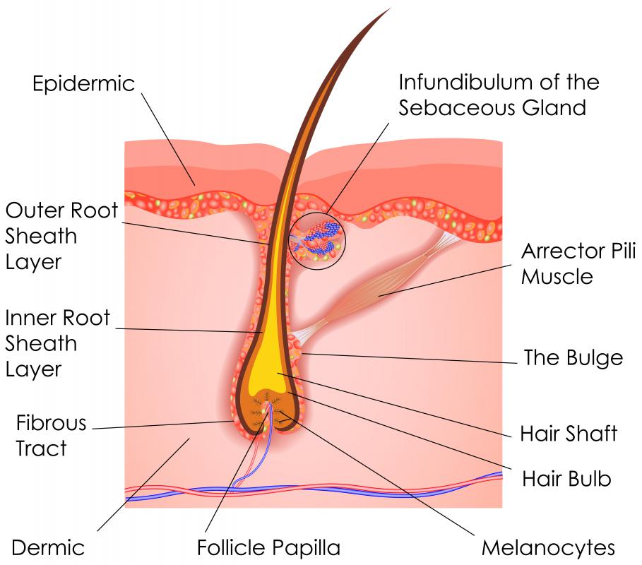


HAIR FOLLICLES 

It is a structure of skin from where the hair grows. The hair follicles are all over the body except the lips, palms, and soles. 

The hair follicle has oe or two sebaceous glands which secrete sebum and lubricate the hair follicles. Apocrine glands help in lubricating the hair follicles of the armpits. 

The hair follicle also consists of a errector pilli which is responsible to create goosebumps when contracted. 

The papilla of the hair follicles consist of epithelial cells responsible fr hair growth and melanocytes which are responsible to impart colour to te hair. 


NAILS 

Nails are known as germinal matrix , a tissue lying beneath the skin behind the fingernail.
The matrix consists of nerves and blood vessels and supplied with the nutrients. 
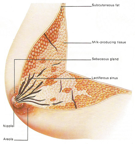


MAMMARY GLANDS

Mammary word comes from the word mammals. The mammary glands are the extensions of the apocrine sweat glands. Mammary glands develop in the females at the time of puberty under the influence of a female sex hormone estrogen. 

The mammary glands are responsible for the secretion of milk for the new born offspring. 

The milk oozes out of the breasts under the influence of hormones prolactin and oxytocin. 


Derivatives of skin 


Wednesday 1 June 2016

understanding the importance of breathing every second

RESPIRATORY SYSTEM 


Respiration is a metabolic process common to all living things. It is a biochemical in which the cells of an organism obtain energy by combining oxygen and glucose, resulting in the release of carbon dioxide, water and ATP. 

PARTS OF RESPIRATORY SYSTEM


  The respiratory system consists of nose, pharynx, larynx, trachea, bronchi and finally the lungs. These are together known as air passages. 

1. The Nose -  The nose consists of an external pair of nostrils which is separated by a cartilage known as nasal septum. 

The nostrils open into the nasal chambers. 

The inner part of the nostrils consists of a lining of mucous and hair.

The nasal hair entraps the dust particles and the mucous tra4ps the bacteria and lubricates the nasal tract.

2. The Pharynx -  The nasal chambers open at the back into a wide cavity, the pharynx, situated at the back of the mouth.

It is a common passage for air and food. 

It leads ito an air tube.

The trachea and a food tube located behind the trachea.

The entrance to the trachea is guarded by a flap called epiglottis which closes it at the time of swallowing food. 

Incomplete closure of epiglottis during swallowing causes cough. 

3. The Larynx - The larynx or the voice box is a hollow cartilagenous structure located at the start of windpipe. The larynx consists of vocal cords which produces sound. 

4. The Trachea - The trachea or wind pipe emerges from the larynx down below in the neck where it is partly covered by the thyroid gland . The walls of the trachea are strengthened by C-shaped rings of cartilage .

5. The Bronchi -   The trachea divides into two tubes , called the bronchi , which enter the respective lungs. On entering the lungs each bronchus divides into fine secondary bronchi, which further divides into still finer tertiary bronchi. 
Bronchioles are the subsequent still finer tubes of tertiary bronchi. By repeated branching , the bronchioles ultimately end in a cluster of tiny air chambers called the air sacs or alveoli.

6. The Lungs -  The lungs are a pair of spongy and elastic organs formed by the air sacs, their connecting bronchioles, blood vessels, etc. The two lungs are roughly cone - shaped , tapering at the top and broad at the bottom. The left lung is slightly smaller to accomodate the heart  in between .


PARTS OF RESPIRATORY SYSTEM ...... 


BREATHING- RESPIRATORY CYCLE 

The respiratory cycle consists of inspiration , expiration, and a very short respiratory pause. 
In normal adults, the breathing rate is 12-18 breaths/min. 

1. INSPIRATION - It results in the increase of the chest cavity which increases the action of the ribs and diaphragm. 

The ribs are moved upward and outward, enlarging the chest cavity. 

Diaphragm is a muscular tissue, which normally remains arched upward like a dome, towards the base of lungs. 

On contraction, it falls or flattens from dome- shaped outline of an almost horizontal plane and thus contributes, to the enlargement of the chest cavity. 

As the diaphragm flattens, it presses the organ inside the and with the abdominal mucles relaxed , the abdominal wall moves outwards. 


2. EXPIRATION -    It is a result of reverse movements of the ribs and diaphragm. 

The external muscles relax and the ribs automatically get depressed . The diaphragm is relaxed and is moved upwards to its dome like outline . The cavity of the thorax is diminished and the lungs are compressed, forcing the air out into the atmosphere . 


RESPIRATORY SYSTEM DISORDERS -   

1. ASTHMA- Asthma is an inflammatory disease of the air ways associated with episodes of reversible over-reactivity of the airway smooth muscle 

The mucous membrane and muscle layers of the bronchi become thickened and the mucous glands enlarge reducing airflow in the lower respiratory tract. 


2. SILICOSIS- This may be caused by longterm exposure to dust containing silicon compounds.

High risk industries are quarrying, granite, slate, sandstone, mining, stone masonry, sand blasting, and glass and pottery work.

Silicosis appears to redispose to the development of tuberculosis, which rapidly progresses to tubercular bronchopnuemonia and possibly military TB. 

Gradual destruction of lung tissue leads in progressive reduction in pulmonary function, pulmonary hypertension and heart failure.


3.ASBESTOSIS - Asbestosis, caused by inhaling asbestos fibres, usually develops after 10 to 20 years exposure, but sometimes after only 2 years. Asbestos miners and workers involved in making and using products containing asbestos are at risk. 


4. EMPHYSEMA - Emphysems is a long term, progressive disease of the lungs primarily causes shortness of breath due to over-inflation of the alveoli or air sacs of lungs. 


5. CHRONIC OBSTRUCTIVE PULMONARY DISEASE - It is an obstructive lung disease because air flow is reduced or stopped. When the air sacs are damaged,, the airways collapse.
This makes it difficult for the lungs to empty the air and th air is trapped in the alveoli. 


5. Lung Cancer - Lung cancer is the uncontrolled growth of abnormal cells that start off in one or both lungs; usually in the cells that line the air passages. The abnormal cells do not develop into healthy lung tissue, they divide rapidly and form tumors.


Effects of smoking on your body ..................


Sunday 3 April 2016

EXCRETORY SYSTEM 

The Excretory system plays a major role in the removal of waste from the body in the form of urea. 
The human excretory system is situated in the dorsal side of the pelvic cavity .

PARTS OF EXCRETORY SYSTEM 

The excretory system consists of a pair of kidneys. The kidney is red bean shaped organ which consists of small units known as the nephrons, which play a major role in the filtration of water and formation of urine. The longitudinal section of kidney consists of a dotted cortex and lighter medulla. 
The outer cortex continues into inner medulla. 
The Tranverse section of the kidney shows the unit of kidney known as nephron. 

The nephron consists of a cup shaped Bowman's Capsule consists of a network of blood vessels and capillaries known as Glomerulus. The Bowman's capsule continues into a tubular convoluted tubule, Proximal convoluted tubule . The The Proximal convoluted tubule continues as ascending limb, loop of Henle, Descending limb, Distal convoluted tubule and collecting tubule. 

A network of connective tubule continues into pelvis which is formed by the union of calyces and renal pyramids present in the renal medulla. 
The tip of the kidney is known as the Hilus, the place from where the pelvis arises and continues into a narrow tube called Ureter.  The Renal Hilus consists of a renal vein and a renal artery , which supplies blood to the kidney. 


Structure of Nephron (Transverse Section of kidney ) 


Location of kidneys 

Ureter is a narrow tube like structure which carries the filtered urine and enters into a bag like structure known as the Urinary Bladder.

Urinary Bladder is a muscular bag like structure which has the capacity for storage of one liter of urine. Once the bladder is full, the bladder opens up the sphincters and gives rise to urination , right through the urethra ad out of the body from the urinary orifice (urinary opening ) .


Parts of excretory  system 


PROCESSES OF KIDNEY 

There are four basic processes in the formation of urine starting with the plasma .


  1. FILTRATION  - It is the mass movement of water and solid substances from plasma to renal tubule that occurs in the renal corpuscle. About 20% of the plasma volume passing through the Glomerulus at any given time is filtered. Around 180 Litres of fluid are filtered by kidneys everyday. Thus, about 3 litres of plasma is filtered 60 times a day.

      2. REABSORPTION - it is the movement of water and solutes from the tubule back into the plasma . Bulk reabsorption, which is not under hormonal control, occurs largely in the proximal tubule. Further bulk reabsorption of sodium in the loop of Henle.  Regulated reabsorption, in which hormone control the rate of transport of sodium and water depending on systemic conditions, takes place in the distal tubule and collecting duct.

     
3. SECRETION- Even after filtration has occurred the tubules continue to secrete additional substances into tubular fluid. This enhances the kidney's ability to eliminate certain wates and toxins. 

4. EXCRETION -  Removal of urine out of the body 
parts and function of excretory system 


Process of excretion in humans 

DISEASES OF EXCRETORY SYSTEM 


GLOMERULONEPHRITIS - It is also called glomerular nephritis. In this disease, the glomerulus , which is an intertwined group of capillaries as a part of the nephrons undergoes inflammation. This can cause acute renal failure too and may cause other disorders like fever, diarrhoea, cough, etc.


HORSESHOE KIDNEY- This is a congenital disorder in which the two kidneys of the patient fuse together to form a horseshoe shape while the child was developing in the womb. It is the most common renal fusion anomaly. Horseshoe Kidney may be a reason for kidney stones or kidney cancer too. 


URINARY TRACT INFECTION -  Urinary tract infection is a bacterial infection in any part of a urinary tract. Usually urine does not contain bacteria in it, but if it multiplies in any part of the tract. The most common Escherichia Coli.


KIDNEY STONES-  It is a disorder in which any part of the urinary tract, basically the ureter gets constricted by stones, which are solid concretions formed in the human kidneys from dissolved urinary minerals. It may obstruct proper urine excretion. 


 A video on the formation of renal calculi or                                             kidney stones 


GOUT-   In this disorder, the body builts up excess amount of uric acid as the kidneys fail to process it properly. 


URETHRIS - This is a disease which is characterized by the inflammation of the urethra. It can lead to painful and difficult urination experience. 


Thursday 17 March 2016

DIGESTIVE SYSTEM 

The digestive is a group of organs working together to convert food into energy and basic nutrients to feed the entire body. Food passes through a long tube inside the body known as the alimentary canal or the gastrointestinal tract. The alimentary canal is made up of the oral cavity, pharynx, esophagus, stomach, small intestine and large intestines. 
In addition to the alimentary canal, there are several important accessory organs that help your body to digest food but do not have food pass through them. Accessory organs of the digestive system include the teeth, tongue, salivary glands, liver, gall bladder and pancreas. 
To achieve the goal of providing energy and nutrients to the body, six major functions take place in the the digestive system :

  • Ingestion 
  • Secretion
  • Mixing and movement 
  • Digestion
  • Absorption
  • Excretion 
PARTS OF DIGESTIVE SYSTEM 
1. MOUTH 
The mouth is the beginning of the digestive tract and in fact, digestion starts here when taking the first bite of food chewing breaks the food into pieces that are more easily digested, while saliva mixes with food to begin the process of breaking it down into a form your body can absorb and use.

             2.  ESOPHAGUS

It is located in your throat near your trachea, the esophagus receives food from your mouth when you swallow. By means of a series of muscular contractions called peristalsis, the esophagus delivers food to your stomach.

3. STOMACH 

The stomach is a hollow organ or container that holds food while it is being mixed with enzymes that continue the process of breaking down food into a usable form. 
Cells in the lining of the stomach secrete a strong acid and powerful enzymes that are responsible for the breakdown process. When the contents of stomach are sufficiently processed , they are released into small intestine. 

4. SMALL INTESTINE 

Made up of three segments- The duodenum, jejenum, and ileum- The small intestine is a 22 foot long muscular tube that breaks down food using enzymes released by the pancreas and and bile from the liver. Peristalsis also is at work in this organ, moving food through and mixing it with digestive secretions from the pancreas and liver. The duodenum is largely responsible for absorption of nutrients in the bloodstream.
Contents of the small intestine start out semi-solid and end in a liquid form after passing through the organ. Water, bile enzymes and mucous contribute to the change in consistency. Once the nutrients have been absorbed and the left- over food residue liquid has passed through the small intestine , it then moves on to the large intestine or colon. 

5. PANCREAS

The pancreas secretes digestive enzymes into the duodenum, the first segment of the small intestine. These enzymes breakdown proteins, fats and carbohydrates.
The pancreas also makes insulin, secreting it directly into the bloodstream.
Insulin is the chief hormone for metabolizing sugar. 

6. LIVER

The liver has multiple functions but its main function within the digestive system is to process nutrients absorbed from the small intestine. Bile from the liver secreted into the small intestine also plays an important role in digesting fat. In addition, the liver is the body's chemical factory. It takes the raw materials absorbed by the intestine and makes all the various chemicals the body needs to function. The liver also detoxifies potentially harmful chemicals. It breaks down and secretes many drugs. 

7. GALL BLADDER 

The Gall Bladder stores and concentrates bile, and then releases it into the duodenum to help absorb and digest fats. 

8. COLON (Large Intestine )

The colon is a 6 foot long muscular tube that connects the small intestine to the rectum. The large intestine ia made up of he caecum, the ascending colon, descending colon, and the sigmoid colon which connects to the rectum. The Appendix is a small tube attached to the caecum. The Large intestine is a highly specialized organ that is responsible for processing waste so that emptying the bowels is easy and convenient. Stool or water left over from the digestive process is passed through the colon by means of peristalsis, first in liquid state and ultimately in a solid form. As stool passes through the colon, water is removed. 
Stool or water left over from the digestive process is passed through the colon by means of peristalsis , first liquid state and ultimately solid state.  Stool is stored in the sigmoid colon until a mass movement empties it into the rectum once or twice a day. 
It normally takes about 36 hours for stool to get through the colon. The stool itself is mostly food debris and bacteria. When the descending colon becomes full of stool or faeces. It empties its contents into the rectum to begin the process of elimination. 

9. RECTUM

The rectum is an 8-inch chamber that connects the colon to the anus. It is the rectum's job to receive stool from the colon to let the person know that there is stool from the colon to let the person know that there is stool to be evacuated and to hold the stool until evacuation. 
When anything comes into the rectum, Sensors send a message to the brain. The brain then decides if the rectal contents can be released or not. If they can , the sphincters relax and the rectum contracts, disposing its contents. If the contents cannot be disposed, the sphincter contracts and the rectum accomodates so that sensation temporarily goes away. 

10. ANUS

The anus is the last part of the digestive system. It is a 2 inch long canal consisting of the pelvic floor muscles and the two anal sphincters. The lining of the upper anus is specialized to detect rectal contents. It lets you know whether the contents are liquid, gas or solid. 
The anus is surrounded by sphincter muscles that are important in allowing control of stool.
The pelvic floor muscle creates an angle between the rectum and the anus that stops stool enters the rectum. When we get an urge to go to the bathroom, we rely on our external sphincter to hold the stool until reaching a toilet, where it then releases the contents. 


video on digestive system 

DIGESTION

Digestion refers to the breakdown of food into smaller components that can be absorbed into the bloodstream. This digestion or catabolism is divided into two types – the mechanical digestion of food that occurs in the mouth when it is physically broken up into smaller pieces and the chemical digestion that occurs in the gastrointestinal tract when the food is broken down into small molecules by digestive enzymes.


COMMON DIGESTIVE PROBLEMS 

  1. Constipation 
Constipation is a condition in which you typically have:

  • fewer than three bowel movements a week
  • bowel movements with stools that are hard, dry, and small, making them painful or difficult to pass
Some people think they are constipated if they don’t have a bowel movement every day. However, people can have different bowel movement patterns. Some people may have three bowel movements a day. Other people may only have three bowel movements a week.
Constipation most often lasts for only a short time and is not dangerous. You can take steps to prevent or relieve constipation.

2.                                                              Gall Stones 

Gallstones are hard particles that develop in the gallbladder. The gallbladder is a small, pear-shaped organ located in the upper right abdomen—the area between the chest and hips—below the liver.
Gallstones can range in size from a grain of sand to a golf ball. The gallbladder can develop a single large gallstone, hundreds of tiny stones, or both small and large stones. Gallstones can cause sudden pain in the upper right abdomen. This pain, called a gallbladder attack or biliary colic, occurs when gallstones block the ducts of the biliary tract.

3.                                      Indigestion 

Indigestion, also known as dyspepsia, is a term used to describe one or more symptoms including a feeling of fullness during a meal, uncomfortable fullness after a meal, and burning or pain in the upper abdomen.